
Gene therapy has been proposed as a promising strategy for the treatment of various genetic disorders and acute acquired diseases such as autosomal or X-linked recessive single-gene disorders including Cystic Fibrosis (C.F.), Adenosine Deaminase deficiency (ADA), emphysema, retinitis pigmentosa, sickle cell anemia, phenylketonuria, hemophilia, and Duchenne Muscular Dystrophy (DMD) [1]. Additionally, recent reports suggest potential treatments for some autosomal dominant disorders, polygenic disorders, different forms of cancers, vascular disease, neurodegenerative disorders, and inflammatory conditions, among many others [2]. Despite the success both in vivo and in vitro, one of the major challenges that still require much effort is the efficient delivery of the therapies, as only a fraction of the delivered genetic material effectively reaches the target site [3]. To address this issue some of the current approaches include the use of viral and non-viral vectors as delivery carriers [2, 3]. Viral vectors have been reported to deliver the gene cargoes with superior efficiencies but thus far have failed to comply with all regulatory and biosafety requirements [4]. These limitations have spurred the development of non-viral safer alternatives that induce a lower immune response and can be prepared inexpensively [2]. As a result, over the past few years, non-viral carriers have gained considerable attention, and particularly those based on nanostructured materials [2, 5]. In this regard, there is a large number of available nanomaterials with unique properties that could potentially be utilized as carriers of biological material [5]. Thus far, only a relatively small fraction of them has been tested for delivery purposes due to issues regarding the required complex synthesis and purification protocols, expensive reagents and most importantly, their limited ability to escape endosomes. These are compartments produced by the invagination of the cell membrane, which eventually matures into lysosomes where enzymes break down their contents [6]. This cell protection mechanism has become a significant obstacle for the full implementation of nanovehicles for drug delivery. This is mainly because the costly transported therapeutic molecules remain trapped and eventually are degraded and expelled from the cells [7]. To address this issue, the nanomaterials have been functionalized with cell-penetrating peptides and proteins, which not only help to bypass the cell membrane but in some cases allow endosome escape [6, 8]. In recent contributions, we have interfaced magnetite nanoparticles with the antimicrobial peptide Buforin II and discovered that the obtained nanobioconjugates exhibited potent translocating and endosomal escape abilities in several cell lines [9]. Despite the success, we wanted to extend these capabilities to other families of nanomaterials such that a more ample arsenal of platforms is available for the rational design of cell-penetrating vehicles to address the needs of particular tissues and diseases. A family of nanomaterials with unique physicochemical properties are the ones based on Carbon [10, 11]. In particular, graphene oxide (GO) has recently emerged as an alternative of great interest for gene and drug delivery mainly because of its unique 2D topology, high surface area, and electrical and optical properties [12]. Additionally, GO is relatively easy to synthesize with controlled particle size and new prospects for large-scale production are becoming available almost on a daily basis [13, 14].
Accordingly, in this work we propose to put forward a new family of cell-penetrating based on GO-based nanoplatforms by interfacing GO with potent translocating peptides. This approach will take advantage of already tested and new peptides as well as the unique interactions of GO with the phospholipids of membranes and endosomes. Ideally, the platform should be able to carry a plethora of pharmacological agents but we are particularly interested in delivering gene therapies. GO was synthesized by the modified Hummers’ method through the oxidation of graphite sheets [6]. Briefly, 0.75 g of graphite sheets were mixed with 4.5 g of KMnO4 in a solution of H2SO4 (90 mL) and H3PO4 (10 mL). The mixture was left at 50°C and under continuous stirring at 350 RPM for 12 hours. Next, the mixture was cooled down at 0°C and H2O2 was subsequently added to quench the oxidation reaction. The resulting mixture was then exfoliated to obtain a solid that was centrifuged followed by resuspension in an aqueous solution of HCl and ethanol. Finally, the as-synthesized GO was filtered, lyophilized, and stored at 4ºC until further use. 100 mg of an aqueous suspension of GO were then silanized by adding 2 mL of tetramethylammonium hydroxide (TMAH) 25% (v/v), 50 µL of pure acetic acid and 1 mL of (3-Aminopropyl) triethoxysilane (APTES) 10% (v/v). A scheme of the reaction is shown in Fig 1A. The silanization was to render free amine groups on the surface of GO to further conjugate the peptide molecules. Thermogravimetric analysis (TGA) and Fourier-Transform Infrared spectroscopy were used to characterize the nanoplatform. FTIR analysis showed the distinctive peaks related to the characteristic carboxyl groups of GO (Fig 1B) as well as the Si-O bonds after silanization (Fig 1B). TGA allowed us to estimate a silanization efficiency of about 35%.
Future work will be focused on conjugating Buforin II and evaluating translocation efficiency by conducting uptake experiments in liposomes and various cell lines. Additionally, we will determine endosomal escape via confocal microscopy by labeling the peptide with fluorescent molecules and looking at colocalization with the fluorescent probe lysotracker.
This work provides an avenue for the development of gene delivery systems based on GO-based nanoplatforms. Moreover, by taking advantage of the great qualities in terms of physicochemical, electrical and optical properties of GO, this study might provide novel strategies to overcome limitations commonly faced such as low stability of the translocating biomolecules and endosomal entrapment.
References:
[1] N. Nayerossadat, P. Ali, and T. Maedeh, “Viral and nonviral delivery systems for gene delivery,†Adv. Biomed. Res., vol. 1, no. 1, p. 27, 2012.
[2] D. Ibraheem, A. Elaissari, and H. Fessi, “Gene therapy and DNA delivery systems,†Int. J. Pharm., vol. 459, no. 1–2, pp. 70–83, 2014.
[3] M. M. Mady, “Cationic liposomes as gene delivery system,†African J. Pharm. Pharmacol., vol. 5, no. 17, pp. 2007–2012, Nov. 2011.
[4] J. L. Shirley, Y. P. de Jong, C. Terhorst, and R. W. Herzog, “Immune Responses to Viral Gene Therapy Vectors,†Molecular Therapy, vol. 28, no. 3. Cell Press, pp. 709–722, 04-Mar-2020.
[5] M. Malmsten, “Inorganic nanomaterials as delivery systems for proteins, peptides, DNA, and siRNA,†Curr. Opin. Colloid Interface Sci., vol. 18, no. 5, pp. 468–480, 2013.
[6] N. D. Donahue, H. Acar, and S. Wilhelm, “Concepts of nanoparticle cellular uptake, intracellular trafficking, and kinetics in nanomedicine,†Adv. Drug Deliv. Rev., vol. 143, pp. 68–96, 2019.
[7] H. Hillaireau and P. Couvreur, “Nanocarriers’ entry into the cell: Relevance to drug delivery,†Cellular and Molecular Life Sciences, vol. 66, no. 17. Birkhauser Verlag Basel, pp. 2873–2896, 05-Jun-2009.
[8] E. Yuba et al., “PH-sensitive polymer-liposome-based antigen delivery systems potentiated with interferon-γ gene lipoplex for efficient cancer immunotherapy,†Biomaterials, vol. 67, pp. 214–224, Oct. 2015.
[9] A. Suarez-Arnedo et al., “Novel BUF2-magnetite nanobioconjugates with cell-penetrating abilities,†Int. J. Nanomedicine, vol. Volume 13, pp. 8087–8094, 2018.
[10] J. Sandoval et al., Carbon nanomaterials as pharmaceutic forms for sustained and controlled delivery systems. 2019.
[11] M. N. Hasan, M. Nafiujjaman, and Y.-K. Lee, 2D Nanomaterials for Gene Delivery. Elsevier Inc., 2019.
[12] N. Rahmanian, M. Eskandani, J. Barar, and Y. Omidi, “Recent trends in targeted therapy of cancer using graphene oxide-modified multifunctional nanomedicines,†Journal of Drug Targeting, vol. 25, no. 3. Taylor and Francis Ltd, pp. 202–215, 16-Mar-2017.
[13] D. de Melo-Diogo, R. Lima-Sousa, C. G. Alves, E. C. Costa, R. O. Louro, and I. J. Correia, “Functionalization of graphene family nanomaterials for application in cancer therapy,†Colloids Surfaces B Biointerfaces, vol. 171, no. May, pp. 260–275, 2018.
[14] P. Liu, S. Wang, X. Liu, J. Ding, and W. Zhou, “Platinated graphene oxide: A nanoplatform for efficient gene-chemo combination cancer therapy,†Eur. J. Pharm. Sci., vol. 121, no. March, pp. 319–329, 2018.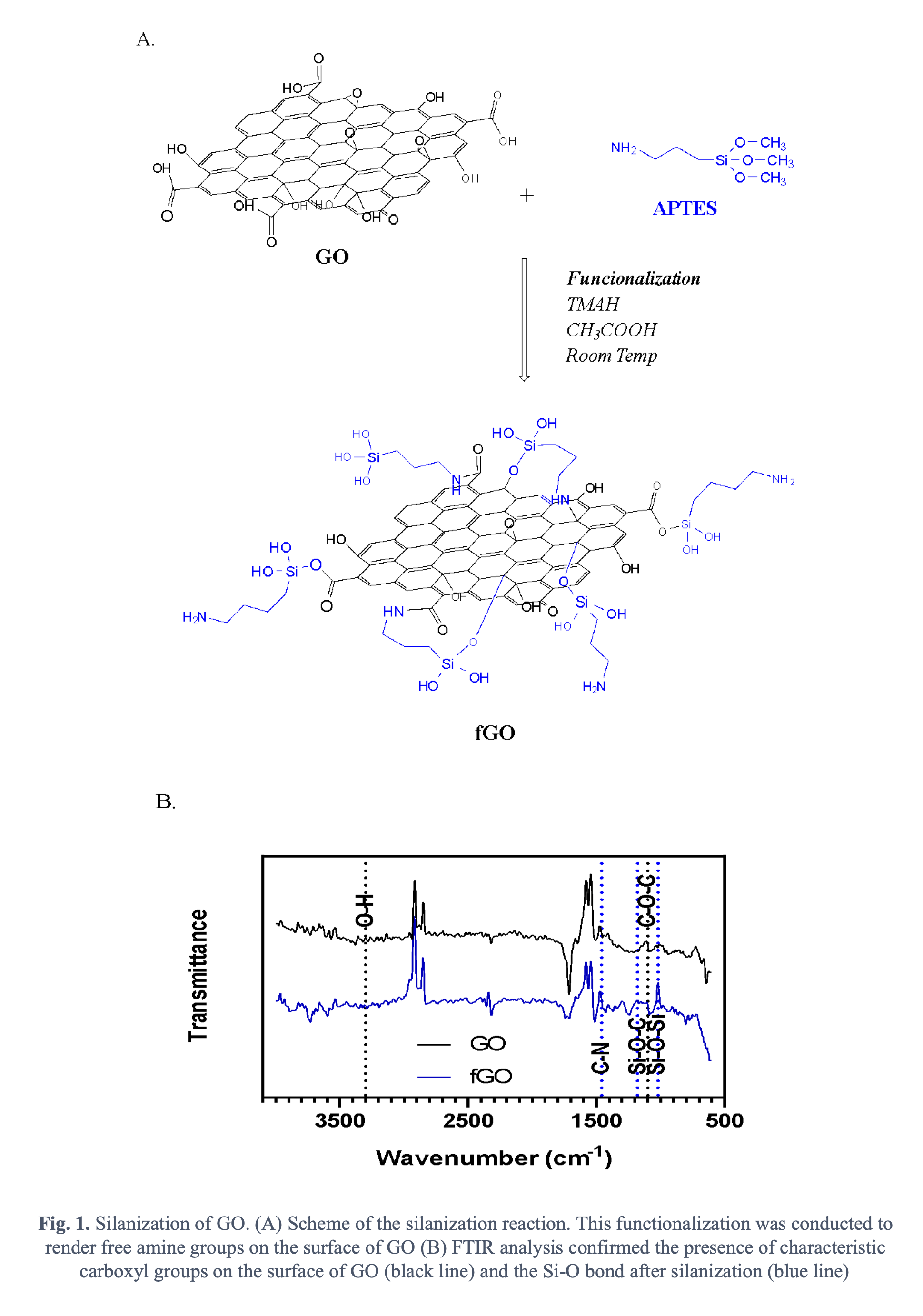
Presenter(s)
Once the content has been viewed and you have attested to it, you will be able to download and print a certificate for PDH credits.
If you have already viewed this content,
please click here
to login.