
Electrospun fiber scaffolds are of interest for ligament regeneration due to their extracellular matrix-mimicking architecture and high surface area to volume ratio. However, limited cell infiltration into these scaffolds has been attributed to tight fiber packing. Consequently, we are interested in combining electrospun fibers with a space-filling hydrogel phase to decrease fiber packing and improve cell infiltration. To this end, we have fabricated a variety of model composites by combining electrospun polycaprolactone/heparin fibers with a collagen hydrogel. The immediate objective of this study was to determine how construction of the composite affects cell viability and morphology, and the longterm objective is to use these models to study cell migration and tissue formation.
In the first set of studies, we prepared thin layers of electrospun fibers on rigid supports to assess how the placement of cells atop a thin collagen gel coat and beneath 0.5 mm and 1.5 mm of collagen gel affects cell viability and morphology. Results indicated that cells attached to and spread along the supported fibers in all cases, but the thicker (1.5 mm collagen) layer diminished cell metabolic activity relative to the other two configurations. We attribute this to diminished availability of nutrients and oxygen. In the second set of studies, thin layers of electrospun fibers were elevated, and the underside filled with collagen gel (to better mimic the mechanical properties of composites). Cells were then seeded directly atop the fibers or combined with collagen and deposited as a 0.5 mm layer gel layer. For the first case cells were able to attach and spread along fibers, while for the second case some cells were able to find and spread on the fibers, while other cells remained rounded up within the collagen layer. Nevertheless, cells within the entire composite remained viable.
Together, these data show that the thickness of the composite can impact cell metabolic activity while the location of the cells (i.e., on the fibers, in the gel) can influence morphology, both of which we anticipate will affect cell proliferation and ligament tissue formation. As a next step, we are currently studying cell migration in response to the chemoattractant FGF-2. Our next step will be to present ligament-inducing morphogens (e.g., GDF-5) within these fiber-hydrogel composites to evaluate their ability to guide tissue formation.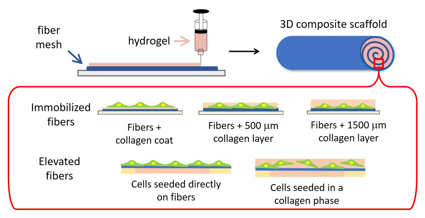
Presenter(s)
Once the content has been viewed and you have attested to it, you will be able to download and print a certificate for PDH credits.
If you have already viewed this content,
please click here
to login.