(15e) Antibody Drug Nanoparticle Induces Synergistic Treatment Efficacies in Breast Cancer Cells
AIChE Annual Meeting
2020
2020 Virtual AIChE Annual Meeting
Food, Pharmaceutical & Bioengineering Division
Delivery of Cancer Therapeutics
Monday, November 16, 2020 - 8:45am to 9:00am
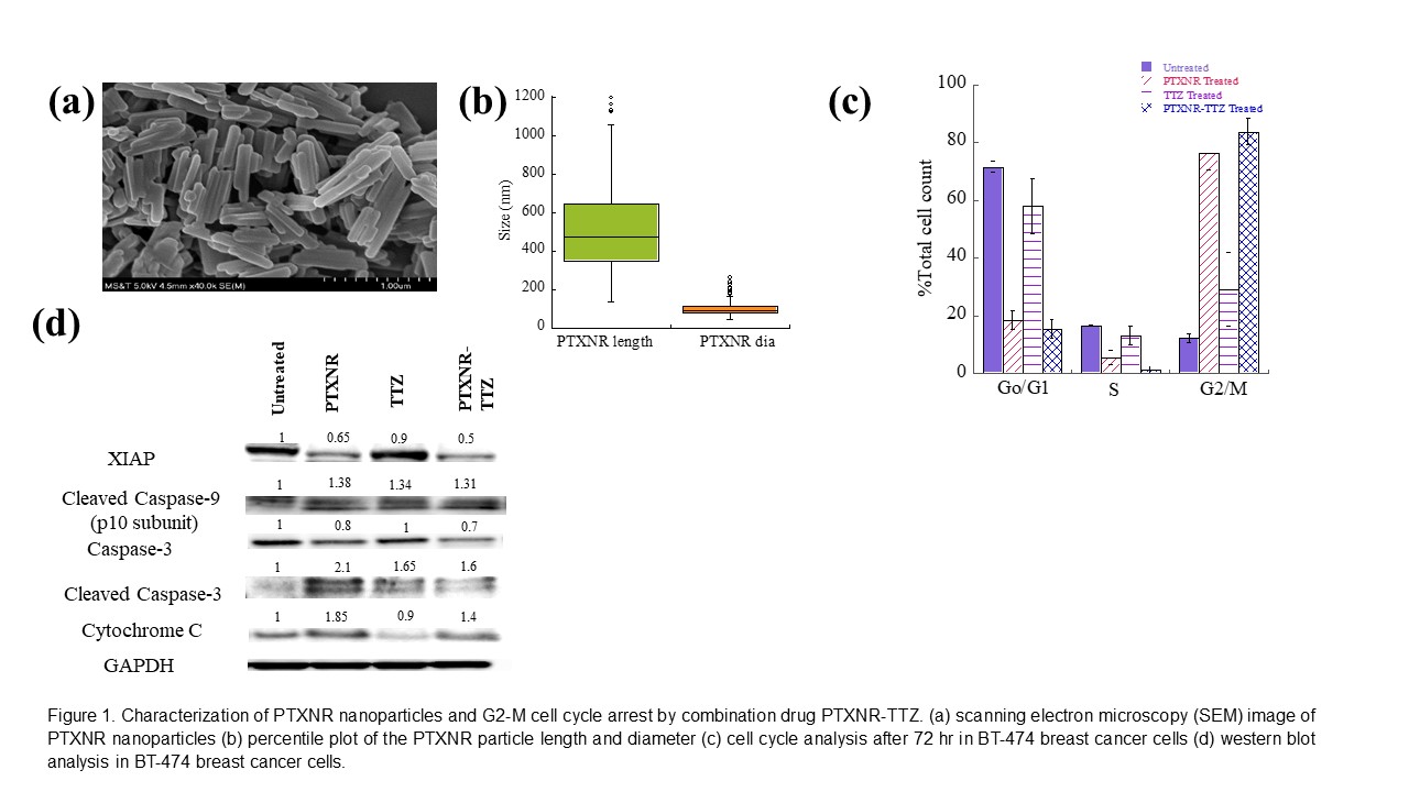
Chemotherapeutic drugs suffer from non-specific binding, undesired toxicity and poor blood circulation which contribute to poor therapeutic efficacy. The goal of this work is to engineer antibody-drug nanoparticles (ADNs) comprising of pure anti-cancer drug nanorods (NRs) in the core with a therapeutic monoclonal antibody on the surface of the nanoparticles for specific targeting and synergistic treatments of cancer cells. The ADNs were designed by first synthesizing ~95 nm diameter × ~500 nm long paclitaxel (PTX) NRs using nano precipitation method (Figure 1a, 1b) . The surface of PTXNRs were functionalized at 2¢ OH nucleophillic site using carbonyldiimidazole and conjugated to Trastuzumab (TTZ) through the lysine group interaction forming PTXNR-TTZ conjugates. The percentage conjugation efficiency was >95% with a drug to antibody mass ratio of 10. In vitro cytotoxicity analysis showed PTXNR-TTZ inhibited >80% of human epidermal growth factor receptor 2 (HER2) positive BT-474 breast cancer cells more efficiently than individual PTX and TTZ treatments alone for 72 h. A combination index analysis indicated synergistic combination of PTXNR-TTZ compared with the doses of each drug alone. The molecular mechansims of PTXNR-TTZ were investigated using cell cycle and Western blot analyses. Cell cycle analysis showed PTXNR-TTZ arrested >80% of BT-474 breast cancer cells in G2/M phase (Figure 1c), while >70% of untreated cells were found in G0/G1 phase indicating that G2/M arrest influenced apoptosis. A similar percentage of G2/M arrested cells were found to induce caspase-dependent apoptosis in PTXNR-TTZ treated BT-474 cells as revealed using Western blot analysis. PTXNR-TTZ treated BT-474 cells showed 1.3, 1.4 and 1.6-fold higher expressions of cleaved caspase-9, cytochrome C and cleaved caspase-3, respectively than untreated cells, indicating up-regulation of caspase dependent apoptotic pathways (Figure 1d).